3D scanning living corals for studying the impacts of microplastics and climate change
Challenge: To quickly 3D scan hundreds of living corals with minimal contact, as part of long-term research studies focused on understanding the physical shape changes taking place in corals due to the effects of microplastics and climate change.
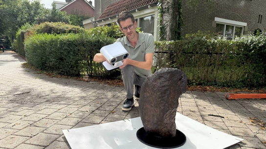
Solution: Artec Spider, Artec Studio
Result: Today researchers can 3D scan an individual coral and obtain its exact physical measurements in under one minute, with submillimeter precision, using the Artec Spider handheld 3D scanner.
Why Artec 3D? The traditional methods of measuring corals are destructive, often killing the coral during the process. But by using the Artec Spider, each coral is scanned dripping wet, then returned to its tank in about a minute’s time without any physical damage to its fragile and sensitive polyps. This means the very same corals survive and thrive for the entire duration of the study, and beyond.

Coral reefs are complex and diverse marine ecosystems providing safe havens around the world for an estimated 7,000–8,000 species of young saltwater fish. With an abundance of food swirling around them, small fish can hide from large predators among a reef’s many crevices and winding passageways.
Despite what many people think, corals are fragile creatures, and very much alive. Each coral consists of hundreds to thousands of little creatures called “polyps.” These polyps are typically anywhere from 1-10 millimeters across, about as thick as a coin, with a soft outside structure, covering their hard, limestone exoskeleton.

Close up of a hood coral (Stylophora pistillata). Image courtesy of Dr. Jessica Reichert
The risk of extinction
Under relentless environmental pressures more than 50% of the world’s coral reefs have died off since the 1950s, due to climate change, overfishing, and pollution. Of those remaining, up to 90% may not survive the next 100 years.
If all the corals die, then so will the fish – and extinction means forever.
Not to mention the widespread impacts that would reverberate around the globe, including the loss of countless millions of fish and other marine life, as well as the dozens of industries that depend upon their constant supply.
Coral reefs have been a vital source of multiple medicines for the treatment of Alzheimer’s disease, cancer, heart disease, etc.
Dr. Jessica Reichert is with a team of researchers at Giessen University* studying the long-term effects of global change stressors on a variety of coral species.

Dr. Jessica Reichert, Artec Spider, and a hood coral (Stylophora pistillata). Image courtesy of Dr. Jessica Reichert
This is made possible by the university’s Ocean2100 facility, which gives researchers the ability to simulate global climate change scenarios and study their effects on corals, reefs, and the organisms living within them.

The Ocean2100 facility at Giessen University. Image courtesy of Dr. Patrick Schubert
Creating the right conditions
With a seawater capacity of nearly 9,000 liters in total, the specially designed aquarium facility consists of 18 separately controlled, 24/7-monitored aquariums holding 265 liters each.
This is where researchers recreate conditions that closely resemble those being faced by coral, such as increased water temperatures, varying levels of acidification, the presence of microplastics, etc.

Various types and sizes of microplastics. Image courtesy of Dr. Jessica Reichert
Reichert and her team are currently working with 30 species of stony corals and over 20 different kinds of other associated reef organisms.
Corals are truly remarkable creatures. Even though individual coral polyps die off, a single coral can theoretically live for hundreds of years. This is why long-term change-over-time studies focused on different coral species are critical. The traditional 60-90 day studies simply are not long enough to measure and understand the physical changes taking place.
Even if studies were longer, a research team’s measurement tools can make or break the accuracy of the results. Colony growth is one of the most important parameters that help scientists study the impacts of stress on corals.

Coral reefs in Bali, Indonesia. Image courtesy of Dr. Jessica Reichert
But with the traditional approaches for determining coral dimensions, using either hot wax or aluminum foil, taking precise, repeated measurements of a single living coral is next to impossible.
Although both methods are still in use today, their limitations are immediately apparent. The hot wax procedure involves dipping a single coral into a tub of scalding paraffin, and then measuring the weight increased by the wax shell once it fully dries.
Unfortunately, the coral never survives this step, and thus subsequent measurements can only be of a separate, neighboring coral, with measurably different dimensions.
While it is possible to use aluminum foil to accomplish a similar yet far less accurate measurement for the coral to actually survive the ordeal, this would entail the researcher moving very slowly for extended amounts of time, producing results nowhere near sensitive enough to quantify a coral’s slow rate of growth (.8 to 5 mm per month).
Introducing new technology
But at Giessen, neither of the older methods have been used for years now. Instead, they’ve fully adopted 3D scanning with the Artec Spider as their measurement modality of choice.

The Artec Spider with a blue coral (Heliopora coerulea) waiting to be scanned. Image courtesy of Dr. Jessica Reichert
With minimal contact, not only do all their coral survive each and every measurement session, but it takes just under a minute for a complete scan.
This has had a profound impact on their productivity. In Reichert’s words, “I usually scan about 50 corals in one afternoon, or 100 in a day. We were never able to work so quickly in the past, not to mention with 100% guarantee of safety for the coral.”
“One long-term scanning project included 300 corals. They had to be scanned closely together, so if I had to do it the old way, without our Spider, it would demand multiple researchers measuring the coral, or having to accept a reduced level of accuracy, because I simply wouldn’t be able to measure them all in such a brief window of time,” Reichert said.
In terms of scanning workflow, Reichert performs all the scans at once, and only later does she process the scans in Artec Studio software.

Scanning a finger coral (Acropora humilis) with Artec Spider. Image courtesy of Dr. Jessica Reichert
Two researchers are key to the scanning, since one will be handling the coral as they’re brought freshly wet from the aquarium and positioned on a rotating table to be scanned, while the other researcher scans with Spider.
They’ve also developed their own ‘toothpicks & needles’ method of positioning certain types of coral, as this allows them to scan the entire coral from top to bottom without having to reposition it even once.
Spider’s ability to scan through moisture has also proven useful. Reichert said, “Even though the corals are wet, Spider just captures them fine, without any problems in terms of reflectivity. We discovered this by experimenting with the scan settings.”
She continued, saying, “Because with Spider we can customize the settings, so, we’re not locked into either being able to scan something or not. This way, what may be impossible to capture with another scanner becomes possible with Spider.”
To give an example of this, when scanning bright, complex corals, they use a higher sensitivity setting, to pick up even the tiniest of intricate structures. Whereas with darker, very simple corals, they switch to a lower sensitivity. Early on, the team drew up a list of optimal scanner settings for each of their coral species.

Screenshots of a Spider scan (without and with texture applied) of a hood coral (S. pistillata). Image courtesy of Dr. Jessica Reichert
After processing the scans in Artec Studio, the 3D models of the coral are typically exported as OBJ files over to MeshLab. They’ve also created their own Python scripts for automatically analyzing the surface area and volume of the coral models.
Other significant possibilities for the Spider scans have opened up as well. A Brazilian colleague, mathematician André R. Backes, developed a C program for analyzing the fractal dimensions of the coral, essentially to see how complex the corals are, as well as how similar they are to one another.
Their latest research has revealed that the rising levels of microplastics in oceans around the world are directly affecting coral growth and health. At the same time, corals are cleaning the waters around them by swallowing microplastics, confusing them for food, and then encapsulating them within their skeletons.

Microplastics embedded within a coral’s calcium carbonate skeleton. Image courtesy of Dr. Jessica Reichert
Greater underwater research
Contrary to what most people think, the biggest sources of microplastics aren’t luxury items such as shower gels and cosmetics. Instead, to a massive degree, the worst offenders are automobile tire dust, synthetic clothing, and paint particles.
Reichert and her team are also investigating the question of whether ingested microplastics are weakening the coral structures and causing them to break more easily. If proven, this will further highlight how coastlines around the world are in peril of losing the only thing standing between themselves and storms and tsunamis: coral reefs.
Apart from the environmental damage, studies have shown microplastics to be neurotoxic, even capable of changing our very DNA.
Referring to these consequences, Reichert said, “We’re looking at the future impacts, what is going to happen in 10 or 20 years if we don’t eliminate or dramatically reduce microplastics right now. We must change course, and we have the power and responsibility to start doing this immediately.”
Another focus of Reichert and her colleagues are the effects of climate change on coral, including warmer water and heightened CO2 levels in the seawater. Particularly ocean acidification, since it strongly affects how the skeleton of the coral is formed.
Also being investigated is whether these morphological changes taking place in coral have any influence on wave actions. Because coral are strongly influenced by the environment around them, if you have a strong current in the water, they will grow in a different way, compared to if there are weak or moderate currents.
In this direction, Reichert’s colleague Juan David Osorio Cano, I.C, M. Ing, PhD, of the Universidad Nacional de Colombia, Sede Caribe, Grupo de Investigación en Oceanografía e Ingeniería Costera (OCEÁNICOS) has been 3D-printing coral models from Reichert’s Spider scans, using them in a flume, to study the effects on water flow around the corals as their shapes change over time.

Using Blender to prepare a 3D model of a hood coral (S. pistillata) for 3D printing. Image courtesy of Dr. Juan David Osorio Cano
By using 3D-printed corals, researchers will have the precise dimensions of the coral, experiment after experiment, so the size and shape variables remain constant, without any risk of coral death due to mishandling.
Reichert said, “I can’t emphasize enough how this research we’re doing just wouldn’t be possible without the 3D scanning we’re doing with Spider. In the past, we were never able to so accurately track these changes in coral growth and shape taking place over time. But now we have the power to do all this.”
She added, “Any researcher with well-trained eyes can look at a coral and see that its shape has changed, but that’s not good enough from a scientific point of view. With our Spider scanner, we can measure this quantitatively, to show exactly how the shape has changed in precisely which ways. Only then can we begin to understand what’s happening and why.”

3D-printed hood coral (S. pistillata) ready for water flow studies. Image courtesy of Dr. Juan David Osorio Cano
Reichert and her team carry on with their work at Giessen, studying many different species of coral.
Yet so much more awaits exploration: the diversity of ways that coral can change shape, which parameters are driving such change, how best to transfer this knowledge up to a reef ecosystem level, then finally understanding how reef complexity will be affected, and what consequences will emerge for the fish living within these reefs, and, ultimately, for us as well.
*Dr. Jessica Reichert is now working at the Hawaiʻi Institute of Marine Biology, in Kaneohe, Hawaiʻi, where her research on corals, together with her use of Artec 3D scanners, continues.
Scanners behind the story
Try out the world's leading handheld 3D scanners.


